Beam shaper for fluorescence microscopy
Reference project
Quantitative analyses in laser-based wide-field fluorescence microscopy
The analysis of measurement images based on laser-based wide-field fluorescence microscopy quickly becomes invalid if it is based on unevenly illuminated Gaussian beam profiles. Using the a|TopShape was able to provide assistance here. A conversion of the Gaussian beams in a microscopy set-up of the CREOL research institute into homogeneous flat-top profiles ensures uniform illumination of the slide and thus more clearly legible images.
Project details
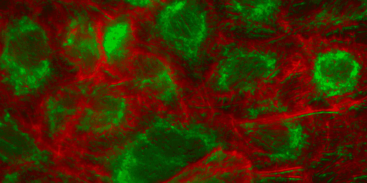
Quantitative analyses in laser-based fluorescence microscopy are made more difficult by uneven illumination of Gaussian profiles. Many factors, such as light source and illumination optics, affect uniformity. These features are particularly challenging when a large FOV (field of view) of the order of several hundred micrometers or millimeters requires investigation. The measurement images are generated in fluorescence microscopy by means of image grids. Individual images are taken in such a way that the edges overlap and they can then be put together in post-processing. If the illumination is not uniform, the final composite image will have darkened edges around each individual image. Measurements of cell and tissue samples thus become unreliable. Another disadvantage of uneven illumination is the uneven activation of the molecules. Those closest to the center of the beam fluoresce more strongly than those on the periphery.
Project realization
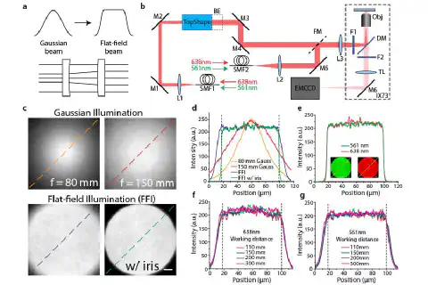
A team of researchers from the College of Optics and Photonics at the University of Central Florida in Orlando, Florida/USA, was able to overcome these problems by using asphericon’s beam shaper a|TopShape and the a|BeamExpander in its microscope set-up (Fig. b). Both products are made of high-precision aspheric lenses, are very compact and have a refractive effect. In this way, it was possible to construct a set-up for flat-field illumination (FFI), which forms the Gaussian beam into a uniform flat-top profile (a). The a|TopShape beam shaper is extremely tolerant to size fluctuations in the incoming laser beam and accepts ± 10%. It works achromatically (e). The long working distance (f) and the high spatial coherence of the FFI enabled an even Epi (illumination and detection from one side of the sample) and TIRF (Total Internal Reflection Fluorescence) illumination for multi-colored single molecule imaging. The unbeatable optical performance of the components used, with a homogeneity > 95%, provides even illumination (c & d) and thus uniform activation of the molecules. In addition, the FFI set-up enables borderless stitched imaging with minimal image overlap (5%).
Experimental characterization of FFI
Figures:
(a) Beam shaping schematic.
(b) Experimental setup. FFI was expanded 1.5x by using the BeamExpander behind the TopShape to provide a full FOV illumination to the sample.
(c) Beam profiles of Gaussian beams collimated by a lens with 80 mm or 150 mm focal length and FFI beams without and with an iris.
(d) Cross-sections taken from beam profiles in (c) along dashed lines. Vertical dashedlines in (d) indicate detection region of camera.
(e) Excitation wavelength dependence of FFI. Cross-sections taken from multicolor images (inserts) with an iris.
(f) Working distance dependence of FFI with 638 nm laser.
Optics for CREOL at a glance:
- a|TopShape and a|BeamExpander enable transformation of the Gaussian beams into a flat top-hat profile and thus uniform illumination of the slide
- Homogeneity of illumination: > 95 %
Further information: I. Khaw, B. Croop, J. Tang, A. Moehl, U. Fuchs, K. Y. Han: „Flat-field illumination for quantitative fluorescence imaging“, In: OPTICS EXPRESS, Vol. 26, No. 12, 11 Jun 2018, pp. 15276-15288
Learn more about the principle of our beam shaper a|TopShape at: asphericon.com/en/solutions/products/beamtuning/beam-shaping/