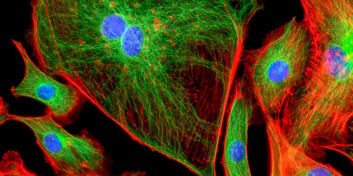
Fluorescence Microscopy
Filters, dichroic beam splitters and beam expanders for cell observation
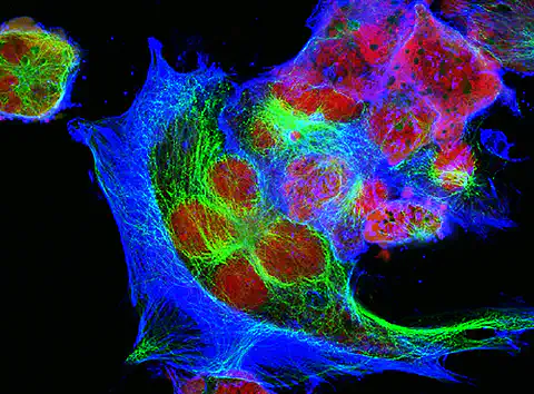
Fluorescence microscopy belongs to the family of light microscopy and is based on the physical effect of fluorescence. The color properties of the so-called fluorochromes are exploited, which are excited by light of a certain wavelength and reflect the absorbed light back again with a different wavelength.
Applications of fluorescence microscopy
Fluorescence microscopy enables morphological investigations, analyses of measured values in the nanometer range and processes of most diverse cultures that become visible in real time. Whether in the field of biochemistry, or medicine: fast and detailed detection of bright, colorful fluorescence facilitates the measuring process of fluorescence microscopy and forms the basis for new findings.
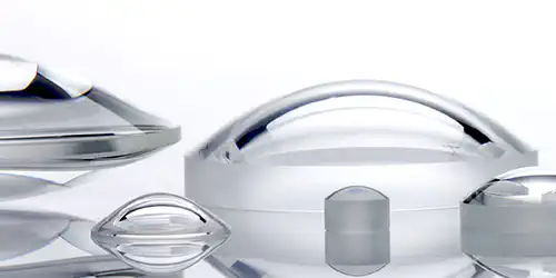
Optimum measurement results and the best resolution require the most precise optics - whether through the optimization and focusing of beam paths, precisely fitting filters or high-quality coatings.
The power of Top-Hat illumination in wide-field fluorescence microscopy
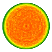
Figure 1: Exemplary wavefront measurement of a Top-Hat profile
Wide-field fluorescence microscopy differs from fluorescence microscopy in its illumination and imaging technique. While traditional fluorescence microscopy illuminates the entire specimen with a broad spectrum of light and collects emitted fluorescence from the entire field, wide-field fluorescence microscopy uses a specific illumination pattern, such as Top-Hat illumination (see Fig. 1), to ensure uniform and controlled light exposure.
This focused illumination allows for higher contrast, reduced photobleaching, and improved image quality, making wide-field fluorescence microscopy particularly advantageous in applications requiring precise and uniform illumination across the field of view. The technique is pivotal in biological, biomedical, and materials research, offering high-resolution visualization of cellular structures and dynamic processes. Its success hinges on illumination quality, affecting data accuracy, sensitivity, and reproducibility.


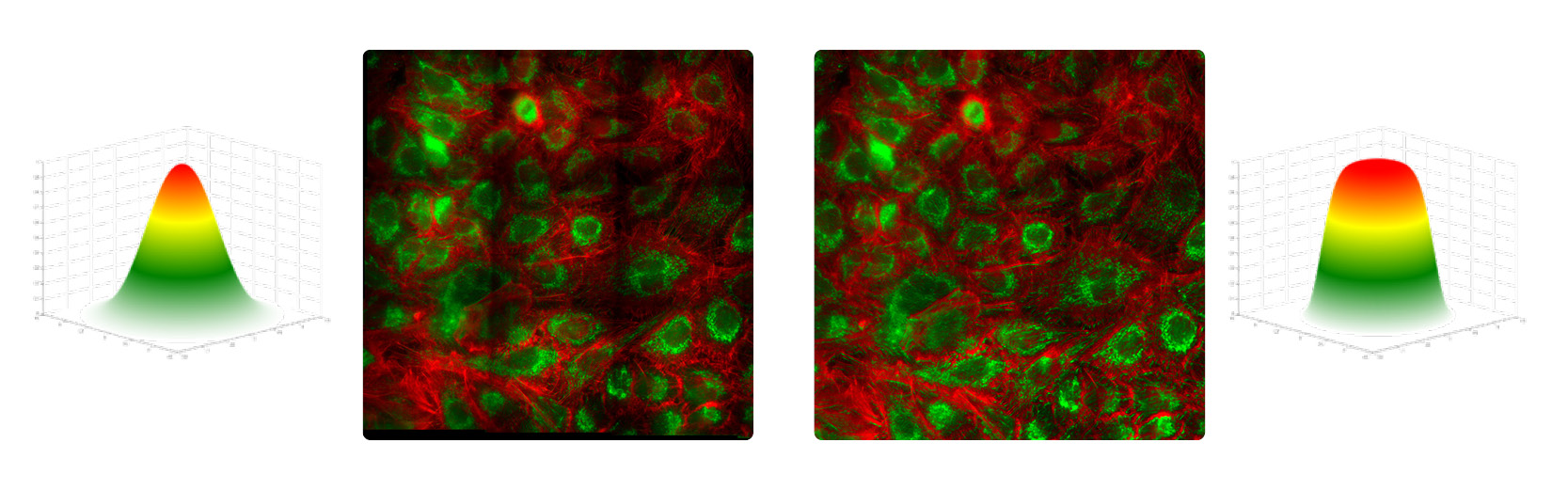
Challenges of Gaussian illumination
While lasers gained popularity for their exceptional properties, Gaussian laser beam profiles present challenges in wide-field applications. The non-uniform intensity distribution (see Fig. 2 left) complicates data analysis and leads to issues like photobleaching and phototoxicity, particularly detrimental in live cell imaging.
Advantages of Top-Hat illumination
Top-Hat illumination, an alternative to Gaussian profiles, provides uniform intensity distribution, minimizing artifacts and enhancing image contrast. This addresses challenges related to photobleaching and phototoxicity, ensuring consistent fluorophore preservation and sample viability. The even light distribution accelerates data acquisition and analysis, streamlining fluorescence microscopy experiments (see Fig. 2 right).
Creating Top-Hat profiles
Several methods exist to create Top-Hat profiles, including truncating the beam, spatial light modulators (SLMs), and beam shaping optics. Truncation, though cost-effective, has drawbacks like power loss. SLMs offer precision but are complex and costly. Beam shaping optics, specifically custom-designed aspheric lenses, provide uniform Top-Hat profiles with minimal optical losses.
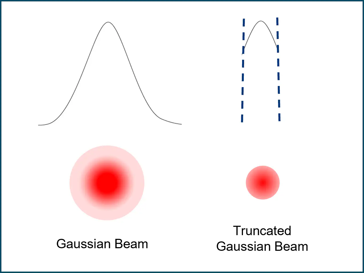
Figure 3a: 'Cutting off' the beam (truncation)
Figure 3b: Sspatial Light Modulator (SLM)
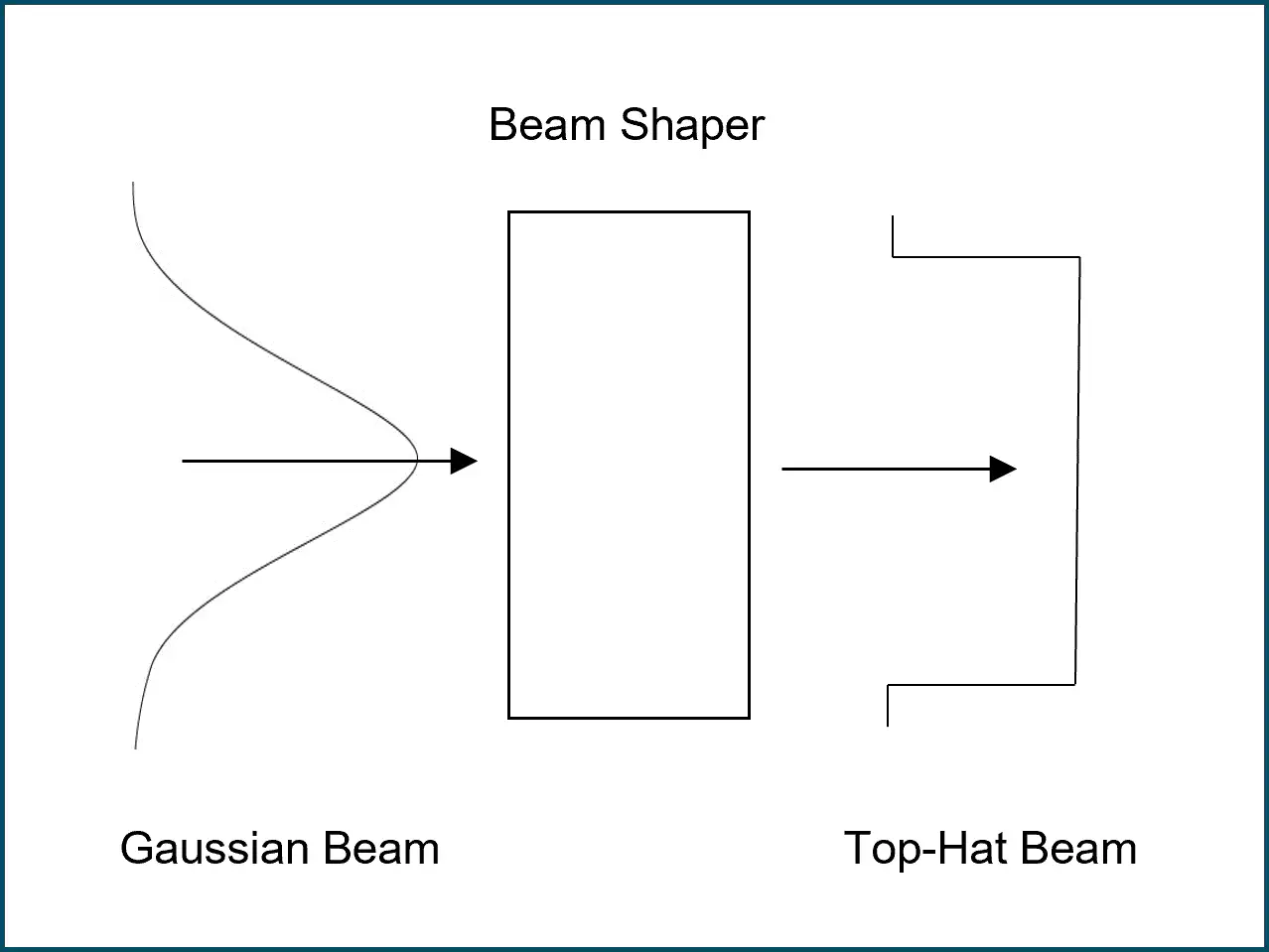
Figure 3c: Beam shaping optics
Application in advanced imaging techniques
Top-Hat illumination significantly benefits advanced techniques like super-resolution microscopy and total internal reflection fluorescence (TIRF) microscopy. In super-resolution microscopy, it enhances localization precision and resolution. In TIRF microscopy, uniform illumination ensures a consistent evanescent field, improving accuracy in studying molecular interactions near cell membranes or interfaces.
asphericons solutions for (wide-field) fluorescence microscopy
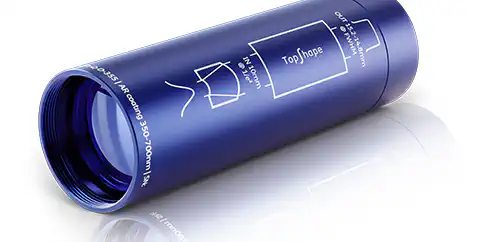
Adopting Top-Hat illumination for wide-field fluorescence microscopy enhances image quality, sample integrity, and experimental efficiency. Aspheric beam shaping solutions, like the a|TopShape, offer a reliable and user-friendly choice for achieving Top-Hat profiles. With our wide range of products, we can offer you a multitude of optimal solutions, from improved imaging qualities through aspheres to a wide variety of filter layers for the separation of different wavelengths


Selected product and service highlights